Anatomical Position and Points-


Spinal Cord-
1.Its long axis is directed vertically downwards.
2.Its lower end is tapering from where filum terminalis starts.
3.Its anteromedian fissure is deeper than posteromedian sulcus.

 .
.


Tongue-
1.Tip of the tongue is directed forwards.
2.Dorsum of the tongue is directed upwards and is covered with papillae.
3.Inferior surface is directed downwards and covered with a smooth mucous membrane.


Muscle of Tongue
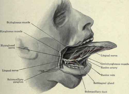
Intrinsic muscle
superior longitudinal -Hypoglossal nerve
transversus -Hypoglossal nerve
inferior longitudinal- Hypoglossal nerve
vertical -Hypoglossal nerve
Extrinsic muscle
genioglossus-Hypoglossal nerve
hyoglossus -Hypoglossal nerve
chondroglossus-Hypoglossal nerve
styloglossus-Hypoglossal nerve
palatoglossus-cranial part of accessory nerve through pharyngeal arch.
All the muscle of the tongue developed from occipital mytome.So there are
supplied by hypoglossal nerve except palatoglossus which is supplied by cranial
part of accessory nerve through pharyngeal arch.
The main functions of extrinsic muscle are altering the tongue's position
allowing for protrusion, retraction, and side-to-side movement.
The main function of the intrinsic muscles is to provide shape and size.
Sensory Supply :
Anterior 2/3rd of the tongue:
General sense : lingual nerve branch of mandibular nerve
Special sense: chorda tympani branch of facial nerve CN VII (carried to
the tongue by the lingual nerve).
Posterior 1/3rd of tongue
General and Special sense: Glossopharyngeal nerve
Extreme Posterior Part :
Developed from the fourth arch so General and Special sense at this part
is supplied by vagus nerve through internal larygngeal nerve.
Lymphatic Drainage of the Tongue

Marginal vessels. These are vessels in the margins of the tongue. They
drain lymphatic vessels to submental nodes, jugulo-omohyoid nodes and the
jugulodigastric node.
Central vessels: These are vessels line central portion of the tongue.
They end in the submandibular lymph nodes and the jugulo-diagastric and the
jugulo-omohyoid nodes.
Dorsal vessels: These run in the dorsum of the tongue. They end in the
jugulodigastric and the jugulo-omohyoid nodes.
Types of papillae

There are four types of papillae
present in the human tongue:
Fungiform papillae - as the
name suggests, these are slightly mushroom-shaped if looked at in longitudinal
section. These are present mostly at the apex (tip) of the tongue, as well as
at the sides. Innervated by facial nerve.
Filiform papillae - these
are thin, long papillae "V"-shaped cones that don't contain taste
buds but are the most numerous. These papillae are mechanical and not involved
in gustation. They are characterized by increased keratinization.
Foliate papillae - these
are ridges and grooves towards the posterior part of the tongue found at the
lateral borders. Innervated by facial nerve (anterior papillae) and glossopharyngeal
nerve (posterior papillae).
Circumvallate papillae - there are only about 10 to 14 of these papillae on most people, and
they are present at the back of the oral part of the tongue. They are arranged
in a circular-shaped row just in front of the sulcus terminalis of the
tongue.
The Taste Area Of Tongue

Structure of taste buds

Each taste bud is flask-like in shape, its broad base resting on the
corium, and its neck opening, the gustatory pore, between the cells of the
epithelium.It covers the tongue ,inferior surface of soft palate,palato glossal
arches,posterior surface of the epiglottis and posterior wall of the
oropharynx.
Formed by two kinds of cells:
a.supporting cells and
b.gustatory cells.
The supporting cells are mostly arranged like the staves of a cask,
and form an outer envelope for the bud. Some, however, are found in the
interior of the bud between the gustatory cell.
The gustatory (taste) cells, a chemoreceptor, occupy the central portion
of the bud which are spindle-shaped, and each possesses a large spherical
nucleus near the middle of the cell.
The peripheral end of the cell terminates at the gustatory pore in a fine
hair filament, the gustatory hair.
The central process passes toward the deep extremity of the bud, and there
ends in single or bifurcated varicosities.
The nerve fibrils after losing their medullary sheaths enter the taste
bud, and end in fine extremities between the gustatory cells; other nerve
fibrils ramify between the supporting cells and terminate in fine extremities;
these, however, are believed to be nerves of ordinary sensation and not gustatory.
The average life of a taste bud is 10 days.


Brain-
1.Superior longitudinal fissure lieas above.
2.Cerebellum lies posterior.
3.Superolateral surface is convex.
4.Medulla oblongata is directed downwards and backwards.
5.Orbital surface is about horizontal.


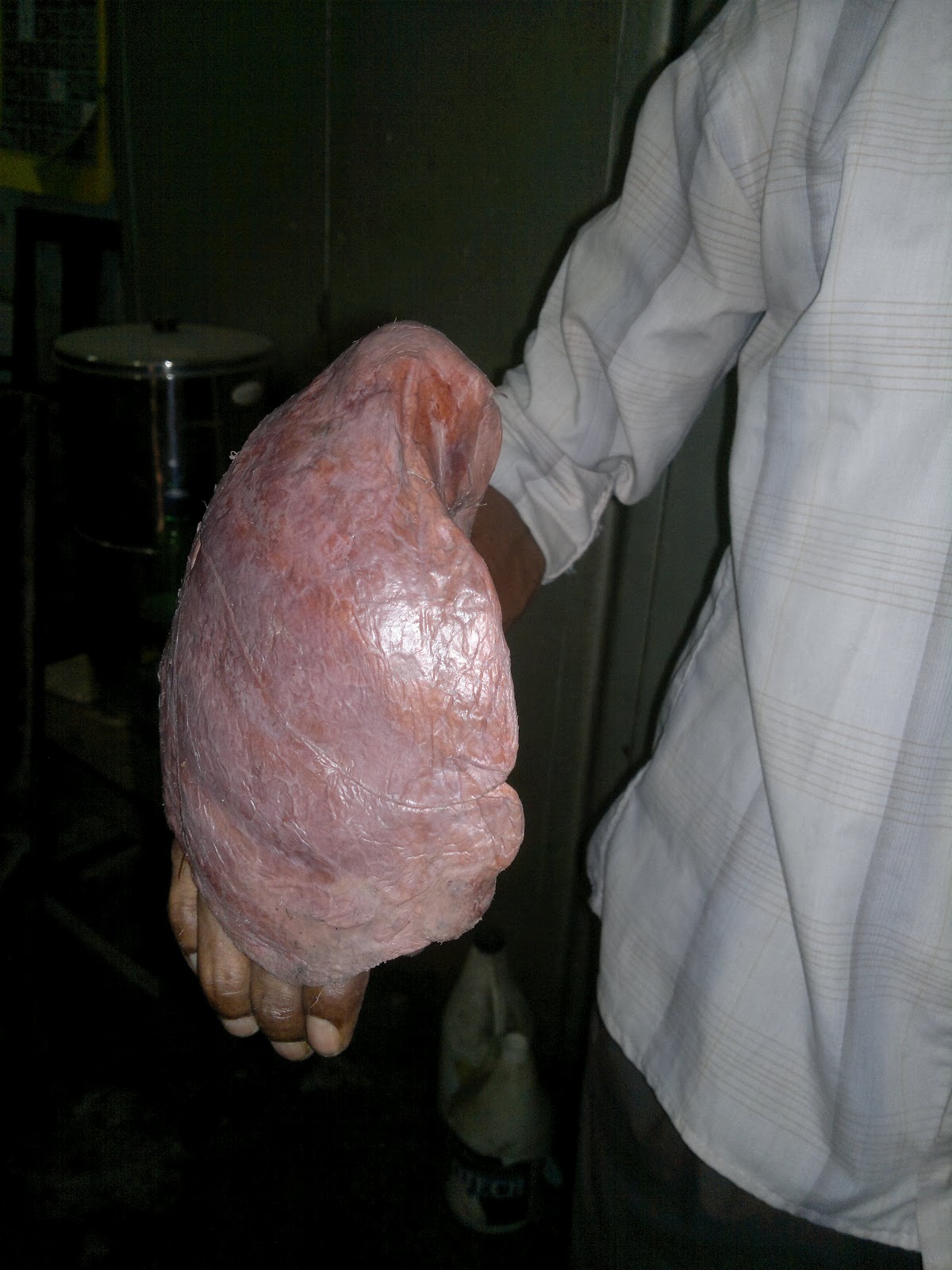


Right Lung-
1.Apex is conical and directed upwards.
2.Anterior border is sharp and is directed forwards.
3.Hilum of the lung lies medially.
4.Base is concave and directed downwards.
5.Hold with right hand


.

Left Lung-
1.Apex is conical and directed upwards.
2.Anterior border is sharp and is directed forwards.
3.Hilum of the lung lies medially.
4.Base is concave and directed downwards.
5.Hold with left hand.


Spleen-
1.The anterior end is expanded and is directed downwards ,forwards and reaches the midaxillary line.
2.The posterior end is rounded and is directed upwards,backwards, and medially.
3.The diaphragmatic surface is convex and smooth.
4.The visceral surface is concave and irregular.
5.The superior border is characteristically notched near the anterior end.
6.The inferior border is rounded.


Liver
1.Groove for the inferior vena cava lies posteriorly and directed vertically downwards.
2.Right lobe is larger than the left and it lies in a higher level than the left lobe.
3.Fossa for gall bladder lies in the inferior surface and this surface is directed downwards,backwards and to the left.


Diaphragm-
1.Pericardium is attached to the central tendon and is directed upwards.
2.Inferior vena cava orifice lies 2 to 3 cm right to the median plane.
3.Oesophageal opening is 2-3 cm left to the median plane.
4.Right dome is slightly higher than the left
5.Aortic opening. is slightly left to the median plane.


Pancreas-
1.The pancreas passes oblique to the left and slightly upwards to left..
2.The anterior surface of the body is concave and is directed forwards and upwards.In the posterior surface ,there is a groove for the splenic vein.
3.The superior border is blunt to the right but narrow and sharp to left and is contact with splenic artery.
4.The head lies within the curvature of duodenum and the tail lies to the left hypochondrium.


Kidney-
1.Long axis of the kidney is directed downwards and laterally.
2.Hilus of the kidney lies medially.
3.Ureter goes directly downwards from the hilus without any twisting.
4.Superior pole lies nearer the median plane then the lower pole.
5.Left kidney is slightly higher than the right kidney.
6.Lateral border is convex and medial border is concave and indented in the middle.
7.Anterior surface is convex and faces antero-laterally.


Heart -
1.Apex is directed downwards ,forwards and to the left.
2.Base lies posteriorly and is directed backwards and to the right.
3.Sternocostal surface is directed forwards,upwards and to the left.
4.Inferior diaphragmatic surface is directed downwards and backwards.


Stomach-
1.The cardiac orifice is situated 2.5cm to the left from the medium plane.
2.The pyloric orifice lies 1.25cm to the right of the median plane.
3.Anterosuperior surface faces forwards and upwards.
4.Posteroinferior surface faces backwards and downwards.
5.The lesser curvature is concave and lies to the right and directed posterosuperiorly..
6.The greater curvature is convex and lies to the left and directed anterolaterally.


Small intestine-
2.Iliac end lies in the right iliac fossa and duodojejunal flexure lies to the left of upper border of second lumbar vertebra.
-Hold the two ends of root of mesentery by two hands obliquely.
-Left end:left to L2 level.
-.Right end:to right iliac fossa.


Large intestine-
2.The concavity of the transverse colon is directed upwards and backwards.
3.The left colic flexure lies at a slightly higher level than the right.


Urinary bladder-
1.Superior surface is triangular and covered with peritoneum.
2.Apex is directed forwards and a neck is directed downwards.
3.Fundus or base is directed backwards.
4.Neck of the bladder directed downwards.


1.Dorsal surface of penis is directed forward.
2.Ventral surface of penis is directed backward.
3.Glans penis is directed downward.


Testis-
1.Superior pole is directed anterolaterally.
2.Sinus of epididymis is directed posterolaterally.
3.Summit of the testis is covered by the head of the epididymis.
4.Anterior border is convex and directed forwards and downwards.


1.Long axis of the vulva/pudendal cleft is directed forwards and downwards.
2.Clitoris lies superiorly and faces anteriorly.
3.Vagina is directed backward and upward.
4.Mons pubis lies infront.
5.Vaginal orifice lies behind and the urethral orifice.
6.Anus lies behind and below.

Abdominal Aorta-
1.Lies vertical slightly left to the midline.
2.Lower end is terminated into right and left common iliac arteries.
3.Coeliac trunk lies anteriorly.

Rectum and Anal Canal-
1.It lies in the median plane.
2.First,it descends downwards and backwards.
3.Second,it descends downwards.
4.Finally,it descends downwards and forwards.
5.Anal canal directed downwards and backwards.
What are the common question that lecturer will ask during
soft part(viscera):
1..Anal canal and rectum:
-How to differentiate whether the anal canal is belongs to a
male or female?
-Parts of anal canal.
-Types of hemorrhoids
-What’s the different between internal and external
hemorrhoid
-Which one is more painful?
Time to time,I will try to update new types of question that
frequently asked during viva...


