The
Identifying Points For Histological Slides.
Identification
of Histology Slide
1.Loose Connective Tissue
-bundle of
collagen and elastic fibre present
-connective
tissue cells are present.
-ground
substance and different connective tissue cells are scattered in the fibres or
in the masses.
2.Hyaline Cartilage
-homogeneous
matrix (glass like)
-cells(chondrocytes)lie
in lacunae forming groups or colony
-dense
fibrous connective tissue covering(perichondrium) present.
3.Elastic Cartilage
-elastic fibres
are abundant.
-elastic
firbres form branching and anastomosing network
-cells are
scattered within fibres
4.FibroCartilage
-coarse
,long ,parallel ,wavy bundle of collagen fibres present
-cells lie
in row between collagen fibre bundles
5.Compact Bone
-presence
of Haversian canal containing neuro-vascular bundle.
-concentric
bony lamellae surrounding central canal present
-lacunae
(containing osteocytes )and canaliculi present.
6.Spinal Cord
-inner ‘H’
shaped gray matter,outer white matter.
-Central canal
present,lined by simple ciliated columnar epithelium(ependyma).
-anterior
median fissure and posterior median sulcus or septa present
7.Cerebellum
-outer gray
matter ,inner white matter present
-cortex
have three layers
-Purkinje
cells in the middle layer of cortex
8.Cerebrum
-composed
of six layers of nerve cells
-pyramidal
cells maybe seen in 3rd and 5th
layers
-outer gray
matter,and inner white matter present
9.Skeletal Muscle
-prominent
transverse cross striation present
-multiple
nuclei located peripherally
-muscle
fibres are cylindrical shaped
10.Smooth Muscle
-cross
striation absent
-nucleus is
single and centrally placed
-spindle
shaped muscle cell
11.Cardiac Muscle
-fibres
branch and anastomose with one another.
-intercalated
disc present
-single
,oval,centrally place nucleus
-cross
striation (faint)present
Artery
12.Large (Elastic )Artery
-a series
(40-60) of concentrically arranged elastic fibres are present in tunica media
-tunica
intima is thicker but adventitia is relatively thin
-external
and internal elastic lamina are not prominent
13.Medium Sized (Muscular)Artery
-a series
of (up to 40) circularly arranged smooth muscle cell present in tunica media.
-internal
elastic lamina is very prominent and thrown into wavy folds.
-tunica
intima is not thick
14.Vein
-large
,irregular luman present but wall is thinner
-tunica
adventitia is the thickest coat
-tunica
media is thin
-elastic
lamina is indistinct or absent
15.Lymph Node
-thin
capsule and thin trabeculae are present
-subcapsular lymph sinus present
-presence
of outer cortex and inner medulla
-cortex
shows closely packed lymph nodules ,some having germinal centres.
16.Spleen
-thick
capsule and trbeculae present
-not
differentiated into cortex and medulla
-red pulp
and white pulp present
-white pulp
(malpighian body )has eccentric arteriole
17.Thymus
-composed
of lobules separated by interlobular connective tissue
-lobule has
outer cortex and inner medulla
-medulla
contain Hassall’s corpuscles
-cortex is
dense but without any lymphatic nodules
18.Tonsil(palatine)
-presence
of tonsillar crypts
-crypts are
lined by non keratinized stratified squamous epithelium
-deep to
crypts lie a number of lymph nodules (with germinal centres)
19.Tongue
-papillae
present
-lined by
non keratinized stratified squamous epithelium
-skeletal
muscle in various direction(u can name the intrinsic and extrinsic muscles if
you want)
20.Oesophagus
-lined by
non keratinized stratified squamous epithelium.
-oesophageal
gland present in submucosa.
-muscularis
mucosa is thock (differentiation from vagina)
-subepithelial
papillae are present.
21.Stomach
-lined by
simple columnar epithelium
-gastric
pit present and gastric gland open at its bottom.
-muscular
layer-outer longitudinal ,middle circular and inner oblique
-mucosal
fold(rugae) present
22.Duodenum
-brunner’s gland present in submucosa.
-leaf like
villi present.
-lining
epithelium is simple columnar with striated border.
-muscle
coat-outer longitudinal ,inner circular.
23.Small Intestine (except
duodenum)
-villi is
present and lined by simple columnar epithelium
-lymphatic
nodule present in submucosa.
-muscle
coat-outere longitudinal ,inner circular.
24.Large Intestine
-numerous
goblet cells present in submucosa.
-lining
epithelium is simple columnar.
-lymphatic
nodules present in lamina propria.
25.Vermiform Appendix
-a complete
ring of lymphatic nodule in lamina propria disrupting muscularis mucosa extends
up to submucosa.
-lining
epithelium is simple columnar.
-muscle
coat-outer longitudinal ,inner circular.
26.Parotid Gland
-only
serous alveoli present
-composed
of lobule separated by interlobular connective tissue.
-Ducts are
profuse and prominent
-groups of
adipose tissue are present.
27.Submandibular Gland
-both serous
and mucous alveoli with serous demilune.
-excretory
duct present.
-scattered
adipose tissue present.
28.Sublingual Gland
-mucous
alveoli abundant ,few serous alveoli present.
-interlobular
connective tissue prominent .
-excretory
duct present.
29.Pancreas
-pancreatic
acini present
-Islets of
Langerhans present between the acini.
-centroacinar
cells present in acini.
-composed
of lobule separated by interlobular connective tissue.
-only
serous alveoli present.
30.Liver
-central
vein in hexagonal hepatic lobules.
-hepatocytes
are radially arranged from central vein
towards the periphery.
-portal
triad lies between the lobule containing a branch of hepatic artery,portal vein
and bile ductule.
31.Gallbladder
-mucosa
projects into fold which look like villi.
-lined by
simple columnar epithelium .
-present
three coats:outer adventitia,middle fibromuscular,and inner mucosa.
32.Epiglottis
-consists
of central core of elastic cartilage
-covered
with non-keratinized stratified squamous
epithelium on upper surface ,pseudo-stratified ciliated columnar on lower
surface.
33.Trachea
-lining
epithelium is pseudo-stratified ciliated columnar with goblet cells.
-trachealis
(smooth) muscle present.
-‘c’ shaped
ring of hyaline cartilage present.
-outer fibrous
coat present.
34.Lung
-alveoli is
lined by simple squamous epithelium
-intra-alveolar
septa is thin containing capillary plexus.
-bronchus
contains pieces of hyaline cartilage.
-different
orders of bronchial tree scattered in alveoli (having thin walls broken at many
places)
35.Skin
-lined by
keratinized stratified squamous epithelium .
-composed
of outer epidermis and inner dermis.
-presence
of hair follicle,sweat and sebaceous gland in dermis.
36.Kidney
-composed
of outer cortex and inner medulla.
-cortex
contains renal corpuscles ,proximal and distal convoluted tubule.
-medullary
ray present
37.Ureter
-lined by
transitional epithelium
-wall has
adventitia,muscular and mucous coat
-adventitia
is composed of fibro adipose tissue.
-star shaped
lumen.
38.Urinary Bladder
--lined by
transitional epithelium
-muscle
coat is thick and crisscross
-submucosa
absent.
-lumen is
irregular.
39.Pituitary Gland
-pars
distalis is very cellular and contain three types of cells –chromophobes,acidophils,basophils.
-pars intermedia forms thin sheet of blue staining
cells
-pars
nervosa- presence of many nerve fibres.
40.Thyroid Gland
-composed
of lobule separated by interlobular connective tissue.
-lobule
contains large number of follicles
-follicle
lined by simple cuboidal epithelium containing colloid particle.
41.Adrenal Gland
composed of
outer cortex and inner medulla.
-Cortex
has:
a)Outer
zona glomerulosa(rounded groups of cells)
b)middle
zona fasciculate (cells in longitudinal column)
c)inner
zona reticularis(cells in anastomosing cord)
-Medulla
contain basophilic cells around the sinusoid .
42.Testis
-seminiferous
tubule present separated by connective tissue.
-between
the tubules interstitial cell of Leydig present.
-tubules
lined spermatogenic cells.
-sperms may
be seen in the lumen.
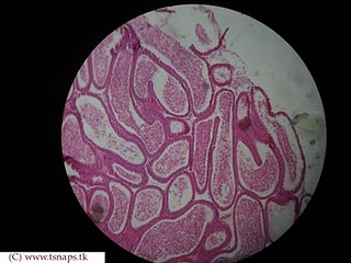
43.Epididymis
-lined by pseudo-stratified columnar with
stereocilia.
-clumps of
sperm in lumen
-lumen is
large
44.Vas Deferens
-thick
middle vascular coat
-lined by
simple columnar epithelium
-muscle
coat-outer and inner longitudinal ,middle circular.
-very thick
wall with small lumen
45.Seminal Vesicle
-mucous
coat shows profuse branching and anastomosis giving it a honeycombed
appearance.
-linning epithelium
is pseudo-stratified columnar.
-outer
fibrous and middle muscular coat.
46.Prostate
-fibromusculogland
organ
-follicle
with wide lumen ,some showing in folding .
-follicle
lined by pseudo-stratified columnar epithelium.
47.Ovary
-lined by
simple cuboidal epithelium
-composed
of outer cortex and inner medulla
-cortex
contain follicles of different stages.
-cortex
also contain corpus luteum.
48.Uterine Tube
-numerous
mucosal fold present ,which almost fill the lumen of the tube.
-mucosa
lined by simple columnar epithelium ,partly ciliated and partly secretory .
-muscle
coat composed of outer longitudinal and inner circular.
-serous is
lined by mesothelium
49.Uterus
-very thick
muscular coat.
-mucosa is
lined by simple columnar epithelium
-uterine
glands are present in lamina propria.
Uterine Gland
They give a
saw –toothed appearance in longitudinal section.
-straight
in follicular phase
-tortuous
in luteal phase
50.Vagina
-lined by
non-keratinized stratified squamous epithelium.
-lymphatic
nodule present in lamina propria.
-muscularis
mucosa absent (differs from oesophagus )
-mucosa has
transverse fold.
51.Mammary Gland
-composed
of lobule of glandular tissue and varying amount of fat.
-lobule are
packed with alveoli and lined by simple cuboidal epithelium .
-duct
present.
[Difference
between resting and active(lactating) period:During resting period –abundant fibrous
tissue,few acini are seen ,but in active stage-fibrous tissue are minimal
,acini abundant.]
52.Umbilical Cord
-two
umbilical artery and one umbilical vein present.
-wharton’s
jelly present..
By Lakdheswaran


.jpg)